16.10.2026 15:51
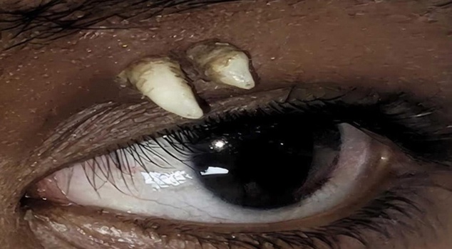
In India, a fully developed canine tooth was surgically removed from the eye socket of a 16-year-old child who went to the hospital with a complaint of swelling in the left eyelid; the case, which entered the medical literature, was diagnosed as an "ectopic tooth."
A fully developed canine tooth was detected in the eye socket of a 16-year-old boy who was admitted to a hospital in Madurai, India, with a complaint of painless swelling on his upper left eyelid. The tooth, removed in a successful surgery performed at Aravind Eye Hospital, was announced to be the first and only case seen in the upper part of the eye socket in the literature.
THE SWELLING ON THE EYELID TURNED OUT TO BE A TOOTH
During the first examination of the young patient, who had noticed a painless swelling on his eyelid about a month before applying to the hospital, two hard and white protrusions were seen. In the Computed Tomography (CT) scan taken, it was determined that these protrusions belonged to a canine tooth and that the crown and root structure of the tooth developed directly from the bone of the eye socket.
THE FIRST "ECTOPIC TOOTH" CASE IN THE LITERATURE
It was understood that the structure removed by surgery was not a "teratoma" type tumor piece that could contain hair, bone, and tooth tissue. Dr. Meghana Tanwar, who evaluated the case diagnosed with "ectopic tooth," defined as the tooth developing outside the place where it should be found, said, "To our knowledge, this is the first and only reported case of ectopic tooth in the upper part of the eye socket."
It was reported that the likelihood of recurrence of this developmental disorder, usually seen in the maxillary sinus behind the cheekbone, is extremely low and that the patient's health condition is good.